Dermoscopy is a modern method of skin examination used in dermatology for the evaluation of all skin lesions for the purpose of diagnosis, differentiation, and monitoring. This technique allows the dermatologist to observe in detail the structures of the skin that are not visible to the naked eye, with dermoscopy representing an extension of the dermatological clinical examination and a non-invasive imaging method used to increase diagnostic accuracy.
Dermoscopy plays a very important role in early detection of skin cancers, especially melanoma, but is useful for the evaluation of all types of pigmented or non-pigmented lesions.
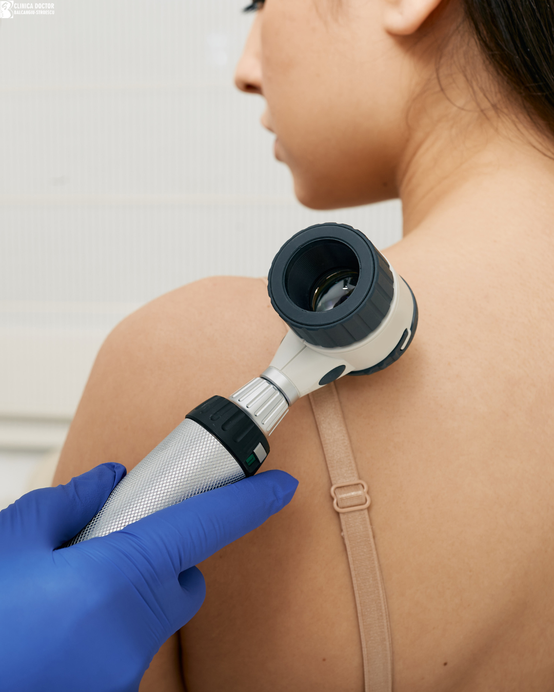
The procedure is quick, painless, and non-invasive, being used routinely in dermatological consultations.
What is dermoscopy?
Dermoscopy is a diagnostic technique that uses a special device called a dermoscope. This is an optical instrument equipped with a source of polarized or non-polarized light and a magnification system, usually 10x, which allow detailed examination of the skin.
Through the dermoscope, the dermatologist can analyze structures at the level of the epidermis and superficial dermis, identifying morphological features that can classify lesions as benign, premalignant, or malignant. The main indications are the evaluation of pigmented nevi and their monitoring over time, early diagnosis of malignant melanoma, and differentiation between various skin lesions (non-melanocytic lesions, vascular lesions, inflammatory/infectious lesions). The dermoscope can be applied to both skin, mucous membranes, and appendages (hair- trichoscopy/nail structures).
How is dermoscopy performed?
The procedure is simple and lasts only a few minutes.
The dermatologist examines the skin using the dermoscope, which is applied directly to the skin surface or very close to it. Sometimes a special liquid (immersion liquid/ultrasound gel/ethyl alcohol/mineral oil) can be used to improve visualization of skin structures.
The examination does not cause pain and does not require special preparation from the patient.
When is dermoscopy recommended?
Dermoscopy is recommended in several situations, not only for diagnosis, but also for screening, differentiation, and monitoring (follow-up), being an essential extension of clinical dermatological examination.
Some people have a higher risk of developing skin cancer and should undergo periodic dermatological and dermoscopic examinations.
These include:
- people with numerous melanocytic nevi (moles): >50- increased risk; >100- high risk
- people with atypical (dysplastic) nevi
- people with personal and family history of melanoma
- people with fair skin (phototype I-III)
- people who have been frequently exposed to the sun or have had sunburns
In these situations, the dermatologist may recommend regular check-ups at intervals of 3/6/12 months.
Additionally, whenever a new pigmented lesion appears or changes in shape, color, size, or structure of a pre-existing lesion are noted, immediate clinical and dermoscopic evaluation is recommended during a dermatological consultation.
Dermoscopy allows early identification of suspicious changes.
Benefits of dermoscopy
Dermoscopy has numerous benefits in evaluating skin lesions.
Among the main advantages are:
- more accurate diagnosis of all skin, mucosal, and appendageal lesions
- early detection of malignant, high-risk lesions
- quick and painless examination
- non-invasive procedure
- monitoring of evolution over time
By using dermoscopy, the dermatologist can decide on the subsequent therapeutic approach for the patient.
Importance of skin monitoring
In addition to dermoscopic check-ups performed during dermatological consultations, periodic self-examination of the skin is also recommended.
People should be alert to:
- appearance of new lesions
- changes in pre-existing lesions
- lesions that grow rapidly, ulcerate/bleed, hurt
- pigmented lesions with unusual appearance
Any change identified through self-examination should be evaluated by a dermatologist as soon as possible.
Conclusion
Dermoscopy represents an indispensable extension of clinical dermatological examination, facilitating detailed analysis of skin lesions in a non-invasive manner. Through its use, diagnosis is significantly improved and early identification of skin cancers is enabled.