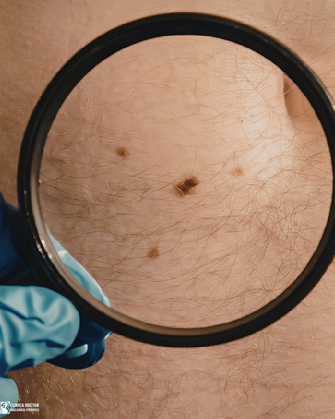
Melanocytic nevi, popularly known as "moles", are frequent pigmented lesions of the skin, present in most individuals. In the majority of cases, they are benign and do not pose a health risk; however, certain changes may suggest the development of suspicious lesions or skin cancer.
In this context, periodic monitoring of nevi and regular dermatological evaluation are essential for early detection of possible pathological transformations. This guide aims to provide relevant information about melanocytic nevi, associated warning signs, and the importance of dermatological examinations.
What are moles
Melanocytic nevi are pigmented skin lesions that appear through the proliferation and accumulation of cells called melanocytes at the epidermal, junctional, or dermal level. Melanocytes are cells that produce melanin, the pigment responsible for skin color.
Nevi may have different clinical characteristics:
- variable color, from light to dark brown
- round or oval shape
- flat or slightly elevated surface
- variable size
Some people have only a few lesions, while others may have a larger number (dozens or hundreds).
Types of melanocytic nevi
There are several types of melanocytic nevi:
· congenital nevi- present at birth or appearing in the first years of life
· acquired nevi- usually appearing in childhood or adolescence; may continue to appear in adulthood
· atypical nevi- lesions with larger size, irregular margins, or varied colors- require dermatological monitoring!
Warning signs – ABCDE rule
Most melanocytic lesions are benign and remain unchanged throughout life. However, certain changes may indicate their transformation into a potentially malignant lesion.
ABCDE rule for identifying suspicious lesions
A – Asymmetry
A normal lesion is usually symmetrical. If one half differs from the other, the lesion may be suspicious.
B – Borders
Benign lesions usually have regular borders. Irregular, jagged, or uneven margins may represent a warning sign.
C – Color
A benign lesion usually has a uniform color. The presence of multiple colors (polychromia) in the same lesion (brown, black, red, or blue) may indicate a suspicious change.
D – Diameter
Nevi with a diameter greater than 6 mm should be evaluated by a dermatologist, especially if they present with other changes.
E – Evolution
Any change in a mole over time is an important signal. Changes may include:
- increase in size
- change in color
- change in shape
- appearance of bleeding or crusts
Other warning signs
Besides the ABCDE criteria, there are other signs that may suggest a suspicious lesion.
These include:
- persistent local itching
- spontaneous bleeding
- appearance of an ulceration that does not heal
- palpation of swellings or nodules in the local region
- rapid change of an existing lesion
- appearance of a new lesion in adulthood
Any of these symptoms requires immediate dermatological evaluation.
Who should have regular dermatological examinations
Periodic dermatological examination is especially recommended for individuals who present with:
- numerous melanocytic lesions on the body
- atypical nevi
- family history of melanoma
- fair skin
- frequent sun exposure or history of sunburns
The dermatologist can recommend the optimal interval for periodic examinations.
Prevention of skin cancers- what we can do
Although not all types of skin cancer can be prevented, certain measures can reduce their risk. Strict photoprotection and periodic self-examination of the skin are the foundations of prevention.
It is recommended to:
- use sunscreen rigorously- daily, with reapplication every 2 hours of sun exposure
- use physical UV protection measures- wearing hats, appropriate clothing, taking shelter under umbrellas and shade
- avoid tanning beds
- periodic self-examination of the skin
How to perform skin self-examination
Skin self-examination is a simple method by which patients can observe possible skin changes. It is recommended to:
- periodic examination of skin on the entire body in front of a mirror- anterior and posterior surfaces of the trunk and limbs, including palms, soles, and scalp
- check less visible areas with the help of a smaller mirror
- observe the appearance of new lesions
Any changes observed should be evaluated as quickly as possible by a dermatologist.
Conclusion
Moles are common and, in most cases, benign. However, monitoring them is important for early detection of possible suspicious lesions.
Periodic dermatological examinations and dermoscopic examination can contribute to early diagnosis of skin cancer.
If you notice changes in a mole or the appearance of a new skin lesion, it is recommended to have a specialist dermatological evaluation.