Moles, medically termed melanocytic nevi, represent frequent pigmentary formations of the skin, resulting from proliferation and accumulation of melanocytes (cells that produce the pigment called melanin, responsible for the natural color of the skin) at the epidermal, junctional, or dermal level. In most cases, they have a benign character and stable evolution, without significant pathological implications.
However, in certain situations, melanocytic nevi may undergo morphological changes suggestive of malignant transformation, especially toward melanoma, the most aggressive form of skin cancer. For this reason, periodic monitoring of pigmented lesions and prompt dermatological evaluation in case of changes are essential for early diagnosis.
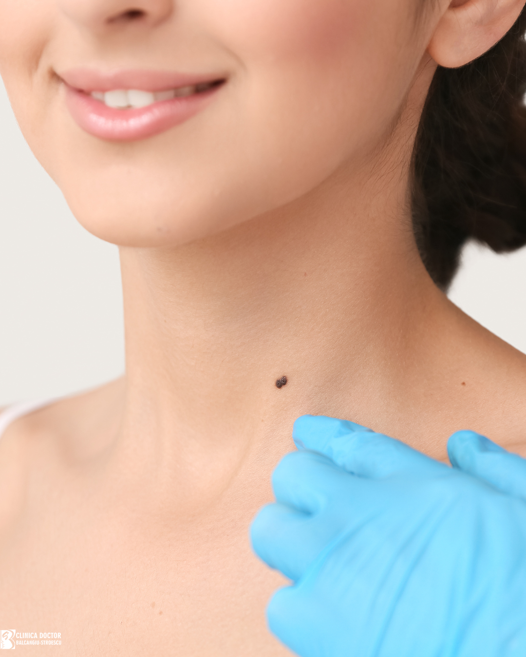
Normal clinical appearance of moles
Melanocytic nevi may have:
- variable color, from light to dark brown
- round or oval shape
- flat or slightly raised surface
The number of these lesions varies considerably among individuals, ranging from a few lesions to hundreds.
When moles can become dangerous
Most melanocytic lesions are benign and remain unchanged throughout life. However, certain changes may indicate their transformation into a malignant lesion.
Melanoma is the most aggressive type of skin cancer and can develop from a pre-existing lesion or as a new lesion.
Early detection of melanoma significantly increases the chances of effective treatment.
The ABCDE rule for identifying suspicious lesions
Dermatologists frequently use the ABCDE rule to identify melanocytic nevi that should be medically evaluated.
A – Asymmetry
A normal lesion is usually symmetric. If one half differs from the other, the lesion may be suspicious.
B – Borders
Benign lesions usually have regular borders. Irregular, jagged, or uneven borders may be a warning sign.
C – Color
A benign lesion usually has uniform color. The presence of multiple colors (polychromy) in the same lesion (brown, black, red, or blue) may indicate a suspicious change.
D – Diameter
Nevi with diameter greater than 6 mm should be evaluated by a dermatologist, especially if they present other changes.
E – Evolution
Any change in a mole over time is an important signal. Changes may include:
- increase in size
- color change
- change in shape
- appearance of bleeding or crusting
Other warning signs
Besides the ABCDE criteria, there are other signs that may suggest a suspicious lesion.
These include:
- persistent local itching
- spontaneous bleeding
- appearance of non-healing ulceration
- palpation of tumefaction or nodules at the regional level
- rapid change of an existing lesion
- appearance of a new lesion in adulthood
Any of these symptoms requires immediate dermatological evaluation.
The importance of dermoscopy
Dermoscopy is a modern method of skin examination used by dermatologists for evaluation of all skin lesions, including melanocytic nevi.
This technique involves skin examination using a special device called a dermoscope, which allows detailed visualization of skin structures.
Dermoscopy helps with:
- early identification of suspicious lesions
- differentiation between benign, premalignant, and malignant lesions
- monitoring their evolution
The procedure is rapid, painless, and non-invasive.
Who should undergo regular dermatological check-ups
Periodic dermatological examination is especially recommended for people who present:
- numerous melanocytic lesions on the body
- atypical nevi
- family history of melanoma
- fair skin
- frequent sun exposure or history of sunburns
In these situations, the dermatologist may recommend regular check-ups to monitor moles.
Prevention of skin cancers- what we can do
Although not all types of skin cancer can be prevented, certain measures can reduce the risk of their occurrence.
It is recommended:
- rigorous use of sun protection- daily, with reapplication every 2 hours of sun exposure
- avoiding excessive sun exposure
- avoiding tanning beds
- periodic self-examination of the skin
Conclusion
In conclusion, although most melanocytic nevi are benign, clinical vigilance, application of evaluation criteria, and use of dermoscopy are essential for early identification of malignant lesions. The ABCDE rule and periodic skin examination can help identify suspicious lesions early. Dermatological consultation remains indispensable in establishing the diagnosis and instituting appropriate therapeutic management.